


Radiofrequency ablation (RFA)
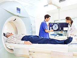
Radiofrequency ablation (RFA) is a minimally invasive interventional radiology procedure that uses heat produced by radio waves to kill abnormal cells. Heat is very effective at eliminating cancerous cells, stopping any further growth. RFA is commonly used as part of a cancer treatment plan. During RFA, imaging technologies, such as ultrasound or CT, help the physician clearly see the affected tissue, and a thin needle equipped with an electrode is applied to the tumor. As controlled currents of energy impact the tissue, the heat generated destroys the cells. Healthy cells grow back in their place. RFA is able to treat malignant or pre-cancerous masses that may be embedded in tissue or in areas otherwise difficult to access.
Why radiofrequency ablation (RFA) is performed
Radiofrequency ablation is considered a complementary treatment for cancer (as opposed to a primary treatment). Patients with multiple small tumors affecting a single organ may also benefit from RFA. It is often recommended when a patient has a small cancerous mass that is creating pain or other physical discomfort. Physicians take several factors into account when determining if RFA is an appropriate treatment option for a patient. Age, previous treatment and response, overall medical status, and cancer stage all play a role in the decision-making process. Cancers in which RFA is commonly utilized include:
-
Breast
-
Bone
-
Kidney
-
Liver
-
Lung
-
Prostate
What to expect during radiofrequency ablation (RFA)
Radiofrequency ablation is typically performed in an outpatient setting with a possible overnight stay for observation. That decision usually depends on the area or organ being ablated and the patient’s current overall medical status. To start, patients are given conscious sedation and local anesthesia is used to numb the area of skin where a small incision will be made. The interventional radiologist guides a long, thin needle with an electrode at the tip through the incision and into the diseased tissue. Imaging guidance helps the provider perform this procedure safely. Once the needle is at the tumor site, radio wave energy is applied to the tumor. It may take several placements on the mass to destroy it completely. The physician will also apply the heat to a margin of healthy tissue to ensure no malignant cells remain. After the treatment, a bandage covers the incision site. The procedure may take two to four hours in total, depending on the size of the tumor, number of tumors, and the organ involved.
Risks and benefits of radiofrequency ablation (RFA)
Radiofrequency ablation (RFA) is generally well tolerated and has high success in stopping the further spread of cancerous cells. In addition to eliminating these malignant cells via heat, the approach also incites an inflammatory response to increase cell destruction. Most patients report very little discomfort following the procedure, which generally does not produce many side effects or complications. However, as RFA is a medical procedure, there is risk. Bleeding, infection at the probe insertion site, and unintended damage to healthy tissue and nearby organs are risks. The imaging guidance used during the procedure helps minimize the likelihood of these issues.
How to prepare for radiofrequency ablation
Patients will likely need routine bloodwork to check their overall immune status and blood clotting ability before radiofrequency ablation. The care team will ask about all current medications and counsel the patient on any that should be paused ahead of the RFA appointment. A team member will also give instructions on when to stop eating and drinking prior to the procedure.
Post radiofrequency ablation
The patient will move to a recovery room after the radiofrequency ablation procedure. Any pain or nausea will be treated during this time, as needed. Patients usually return home after a single night hospital stay. Depending on the level of pain at discharge, prescription pain relief may be ordered. Pain should resolve within a few days. Follow-up CT scans will be scheduled to check the ablation site to ensure that no cancerous cells have returned. If it is found that cancerous cells have returned, another RFA treatment may be recommended.
Microwave ablation (MWA)
Microwave ablation (MWA) is a minimally invasive interventional radiology procedure that uses the heat generated by electromagnetic currents to destroy abnormal cells. The application of heat to tumor cells prevents any further growth or spread. MWA is used regularly for cancer treatment.
During MWA, imaging guidance (ultrasound or CT) visualizes the affected tissue for the interventional radiologist. From here, the doctor safely inserts a needle with an electrode tip through the skin and into the problematic area. The electrode generates heat to eradicate the questionable cells. MWA is effective for eliminating malignant cells, as well as pre-cancerous or injured cells.
Why microwave ablation (MWA) is performed
Microwave ablation is a newer, well-tolerated treatment approach for lung, liver, kidney, prostate, and bone cancers. Heat is highly capable of generating rapid cell death. MWA is typically recommended when a patient is unable to undergo surgical tumor resection (removal) due to advancing age, overall disease burden, or issues with the patient’s heart or pulmonary functioning. MWA might be a sole approach to alleviate cancer symptoms when a mass is too risky to operate on, or it could be part of a broader, multi-therapy treatment plan. The tumor’s core attributes (size, staging, and site) are what helps a provider determine if MWA will be effective in eliminating malignant cells.
What to expect during microwave ablation (MWA)
Microwave ablation is performed in an outpatient setting by an interventional radiologist. However, depending on the tumor site or organ, an overnight stay following the procedure may sometimes be required. Patients are relaxed with conscious sedation prior to the start of the procedure. Next, local anesthesia is applied to the area of the skin that will be accessed with a small incision. A needle with an electrode tip is guided through the incision with imaging guidance in the form of ultrasound or CT. This helps the provider safely access the tumor mass. Once at the tumor, the provider will initiate the microwave energy to agitate the tumor tissue. This agitation ultimately leads to the destruction of diseased cells. Once the procedure is done, usually within two hours, the electrode is removed and the area is bandaged. From here the patient moves to a specialized recovery area or else is admitted to the hospital for an overnight stay.
Risks and benefits of microwave ablation (MWA)
Microwave ablation is a safe procedure with many benefits. Most importantly, the treatment approach has high success in stopping the growth or spread of cancerous cells. MWA also offers patients a rapid recovery time with less pain when compared to surgical tumor management. MWA does carry some small risks, including bleeding, infection, and accidental heat damage to nearby (healthy) tissue. The usage of imaging guidance aims to reduce these risks as much as possible.
How to prepare for microwave ablation (MWA)
Patients will be asked about all current medications, as some may need to be stopped prior to the scheduled procedure. Bloodwork may also be ordered to make sure that the patient has adequate immune system response and that their blood clots normally. The care team will communicate any restrictions on eating and drinking prior to the appointment time.
Post microwave ablation
Any immediate post-procedure pain will be addressed with pain medication while the patient is still at the hospital. Over-the-counter pain relief can treat any residual pain after the patient returns home. Most MWA patients are able to resume normal daily activities within only a few days following the procedure. The vast majority of MWA patients experience no pain at the one-week mark after the ablation.
Our providers
Expert interventional radiology care
Getting the care you need starts with seeing one of our interventional radiologists.