


Robotic cardiac surgery is an innovative approach that incorporates the latest minimally invasive surgical techniques. Combining the agility of robotic instruments with a high-definition 3D camera, Yuji Kawano, MD, the lead surgeon, and the surgical team can perform cardiothoracic procedures through very tiny incisions in the chest wall that would not be possible without the da Vinci robotic technology.
This highly specialized approach can provide patients with enhanced recovery, shorter hospital stays, and fewer complications as compared to conventional methods. Robotic cardiac surgery may be applied to treat aortic, mitral and tricuspid valve conditions, atrial fibrillation, and cardiac tumors, for low-risk surgical patients as well as high-risk patients with complex medical conditions.
Conditions
Aortic Valve Disease
Types of aortic valve disease include:
- Aortic regurgitation: in which blood leaks backward through the valve.
- Aortic stenosis: a narrowing of the valve.
Mitral Valve Disease
Types of mitral valve disease include:
- Mitral regurgitation: in which blood leaks backward through the valve.
- Mitral valve prolapse: in which the valve’s leaflets bulge into the left atrium.
- Mitral stenosis: a narrowing of the valve.
Tricuspid Valve Disease
Tricuspid valve disease includes:
- Tricuspid regurgitation: in which blood flows backward through the valve because the tricuspid valve doesn’t close properly.
- Tricuspid stenosis: a narrowing of the tricuspid valve that limits how much blood flows through the heart.
Cardiac tumors and masses
Tests
Our specialists may recommend one or more diagnostic and imaging procedures.
- Cardiac catheterization: Cardiac catheterization is a minimally invasive way to diagnose and treat a variety of heart and vascular conditions by guiding thin, flexible tubes called catheters through blood vessels to problem areas.
- Chest X-ray: Chest X-rays use a small dose of radiation to create pictures of the structures inside the chest, including the lungs, heart, and chest wall.
- Echocardiogram: An echocardiogram uses high-frequency sound waves to create images of your heart.
- Electrocardiogram (ECG): An electrocardiogram, also known as an ECG, measures the heart’s electrical activity.
- Magnetic resonance imaging (MRI): Magnetic resonance imaging, better known as cardiac MRI, is a combination of radio waves, magnets, and computer technology to create images of your heart and blood vessels.
- Transesophageal echocardiogram (TEE): Transesophageal echocardiogram allows us to take very detailed images of your heart structure from a probe in your esophagus.
Our location
Distance from Change locationEnter your location
MedStar Health Cardiac Surgery at MedStar Washington Hospital Center
110 Irving Street, NW First Floor Washington, DC 20010
202-877-3503

Meet Dr. Yuji Kawano


Patient stories
-
Robotic heart surgery gets Michael back to life

-
Washington Area's First Robotic Heart Surgery Performed at MedStar Washington Hospital Center
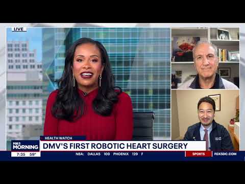
Cardiac surgeon Yuji Kawano, MD, performed the Washington area’s first successful robotic mitral valve surgery at MedStar Washington Hospital Center. Just three days after the robotic heart procedure, patient Brian Cohn was discharged home, thanks to this advanced minimally invasive surgical treatment for his mitral valve. Watch now, as Dr. Kawano and Brian share their experience with Fox 5 DC.
Related services
Ask MHVI
Have questions for our heart and vascular program? Email us at AskMHVI@medstar.net.