


Find care now


If you are experiencing a medical emergency, please call 911 or seek care at an emergency room.
Cardiac PET scan technology promises to elevate cardiovascular diagnoses to a completely new level.
Positron emission tomography (or PET) is traditionally associated with cancer care. Oncology, in fact, is where the technology developed and advanced. But today, this type of scan can be applied to other areas of the body—and it’s becoming a real game-changer in cardiac care.
Most other forms of imaging technology unveil physical or anatomical structures in the body. For example, a traditional X-ray can show broken bones, blockages in the sinuses or disease in the lungs. Mammography uncovers breast disease. CT scans offer detailed imaging in the form of virtual slices of organs that can illustrate abnormalities like tumors and clots. MRI delivers even deeper detail, especially within soft tissue.
But this scan is different. It uses molecular imaging to determine function at the cellular level. In other words, where other imaging methods see physiology, PET sees biology and function.
Other imaging methods see physiology, PET sees biology—function at the cellular level. That’s bringing new accuracy to cardiac screening, says Dr. Carlos Garcia. https://bit.ly/2ZXxF3v via @MedStarWHC
How It Works
Like all living things, cells need food. Most of our cells run on glucose, a type of sugar, and cardiac muscle cells run on fatty acids. In nuclear imaging, we take advantage of that fuel cycle and use these nutrients as a carrier for a radiopharmaceutical, an indicator made with radioactive isotopes. As the cells consume the food, they absorb the radiopharmaceutical, and the camera technology tracks it in real time.
The level of radioactivity needed is relatively low, so the patient is exposed to minimal radiation. And as technology improves, dosages are decreasing. In fact, compared to other nuclear studies, the required dose may be up to two-thirds less. PET is safe and getting safer.
What It’s Best For
When it comes to cardiac imaging, this type of scan is well suited to assess perfusion—how well the coronary arteries supply oxygen to the heart muscle. It can also measure viability, determining which heart muscle cells are healthy and living if a heart attack or other damage has killed cells and created scar tissue. And, lastly, we can use it to assess disease processes infiltrating the heart muscle, especially diseases that cause cardiomyopathy.
Before this new technology was introduced, the gold standard for assessing blood flow was single-photon emission computerized tomography (SPECT). SPECT allows us to identify areas of the heart muscle that are not receiving an adequate blood supply. These will present as perfusion “defects” when the cells do not uptake the radiopharmaceutical. SPECT is still a very useful tool in the cardiologist’s toolbox. But PET does more.
SPECT can deliver equivocal results at times. For example, breast tissue can reduce the amount of activity captured by the camera. Increased body habitus or a buildup of fluid around the lungs, which often accompanies heart disease, can have a similar effect. PET, when combined with CT, can see through all those obstructions and gives the doctor more detailed data.
Measuring Blood Flow
Not only do the scans measure blood flow superior to SPECT, they can accurately measure coronary flow reserve, the body’s ability to maximize blood flow to the heart when oxygen demand rises, such as during exercise.
Oxygen is so critical that the body will adapt to narrowed arteries, rerouting blood through other vessels and even growing new vessels to replace blocked ones. But even with that remarkable response, flow can still be reduced at the capillary level—the tiniest blood vessels where oxygen molecules move from blood to cells. Other cardiac imaging studies cannot see that level of detail and may not show flow reduction at all. With PET, we can tell with great accuracy when cells are threatened by lack of oxygen.
With that level of detail, the technology can deliver a number of miraculous advances. One of the most impactful: the potential for early detection of coronary artery disease, even when no symptoms may be present.
Fighting Cardiomyopathy
This new scanning technology is also proving to be a powerful weapon against infiltrative cardiomyopathy, when foreign cells invade cardiac muscle tissue.
One cause of this cell invasion is sarcoidosis, a challenging autoimmune disease that can affect any organ in the body. Sarcoidosis is a hyper-immune response to inflammation that causes the body to over-produce immune cells. They clump together to form granulomas. In the heart and blood vessels, sarcoidosis can mimic traditional coronary artery disease—and do just as much damage. Once in the heart muscle, sarcoidosis can stiffen the tissue, making it less elastic and reducing pumping efficiency, called cardiomyopathy.
Over time, the invading cells displace healthy cardiac muscle and the heart has a harder time doing its job. This condition can look like full-blown congestive heart failure, with fatigue, shortness of breath, swelling of the extremities and fluid buildup.
Because PET shows what’s happening at the cellular level, we can use it to assess these disorders very accurately and determine treatment and management. As such, it is difficult for any study to be more accurate in identifying these kinds of problems.
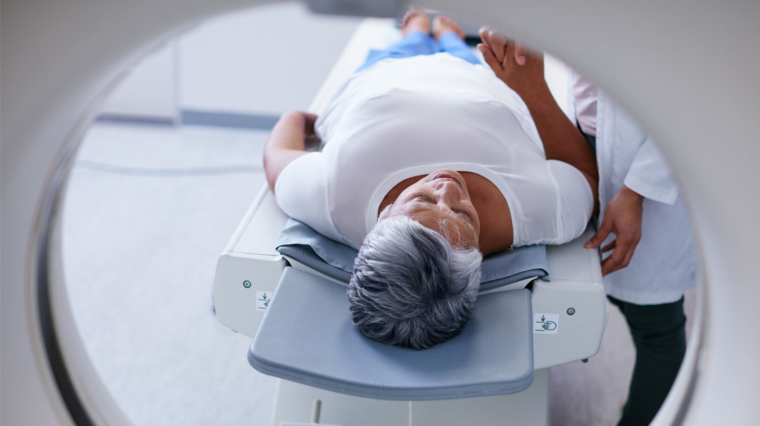
What to Expect From a Scan
Every patient is unique and is asked to do some dietary preparation, depending on the study. Generally, this involves fasting—to make your cells hungry for glucose—or going on a low-carb/high-protein diet to keep the cells sensitive to fatty acids. The prep is very simple, and our staff calls every patient to review the instructions and answer questions.
When you arrive, we’ll start an IV to administer the radiotracer. You’ll have no dietary or exercise restrictions when you go home, but we will ask you to drink extra fluids to flush the tracer out of your system. An average stay is around two hours, including prep, the study and discharge.
Looking Ahead
PET will become even better over time.
The perfusion radiopharmaceuticals we use today are very short-lived, lasting only about 75 seconds. So we cannot, for example, run a scan while the patient is on a treadmill, as we do in a traditional stress test. However, Fluorine-18 Flurpiridaz, a new agent that will likely gain FDA approval next year, will make scanning even more versatile, as it will last up to two hours in the body and enable stress testing. It also binds to mitochondria—the batteries that power every cell—which will help increase the accuracy of the tests. And it will likely cost less than the agents in use today.
The MedStar Washington Hospital Center Difference
At the Hospital Center, our patients have access to state-of-the-art nuclear and molecular technology like cardiac PET—not every medical center offers this technology. Our team of full-time nuclear medicine experts are at the leading edge of this specialty and fully dedicated to it.
We focus on our patients’ peace of mind. We know that being placed inside a large, unfamiliar machine can cause apprehension, and we work hard to make the experience as comfortable as possible.
Our team members take pride in treating every patient like a member of their own family. When you come to the Hospital Center for a nuclear medicine study, you’re not a stranger. We are friendly, welcoming, accessible and reassuring, helping to put every patient at ease.
This is possible thanks to our truly collaborative culture. Each member of our team is like a member of an orchestra. We play our hearts out, doing our absolute best to make the music that will create the wonderful symphony that will be your experience.
And our patients respond well to that.
