


Advanced therapies to reduce blood clots and stroke risk
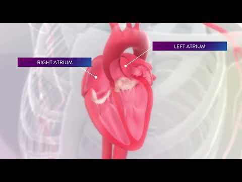
The left atrial appendage (LAA) is a small pocket attached to the left atrium, one of the heart’s four chambers. When atrial fibrillation (AFib) disrupts normal blood flow through the heart, blood can collect and clot within the LAA, increasing the risk of stroke.
The doctors in our Electrophysiology program were among the first in the nation to use the WATCHMAN™ device, a groundbreaking AFib and LAA therapy. We perfected its use through clinical trials, and we continually research and refine new procedures to treat even the most complex arrhythmias.
What to expect during left atrial appendage closure
The WATCHMAN and the AMPLATZER Amulet are the two devices we use to close the LAA.
-
WATCHMAN: Your doctor will place the WATCHMAN device, which is similar to a permanent mesh filter, in the opening of the LAA. Over time, scar tissue will form over it and seal off the pocket. Our team is continuing to participate in clinical trials to improve the use of this device.
-
AMPLATZER Amulet: Currently used in clinical trials, the Amulet device is similar to and smaller than the WATCHMAN and has the potential to benefit a wide range of eligible patients. MedStar Health is the first program in Maryland, Virginia, and Washington, D.C., to offer this device.
Left Atrial Appendage Occluders (LAAOs) Reduce the Risk of Stroke in AFib Patients

Dr. John Wang discusses left atrial appendage occluders for AFib patients. Left atrial appendage occluders seal off the left atrial appendage -- reducing the risk for stroke and eliminating the need for blood thinnners.
Electrophysiology program
We are leaders in developing and using the latest procedures and technologies to treat heart rhythm disorders, and our cardiac electrophysiology laboratory is one of the most sophisticated in North America.
Tests
Chest X-ray
Chest X-rays use a small dose of radiation to create pictures of the structures inside the chest, including the lungs, heart and chest wall.
Echocardiogram
An echocardiogram uses high-frequency sound waves to create images of your heart.
Electrocardiogram (ECG)
An electrocardiogram, also known as an ECG, measures the heart’s electrical activity.
Event Monitors
An event monitor is a small device that records the heart’s electrical activity. It’s similar to an electrocardiogram, but whereas an electrocardiogram takes place over a few minutes, an event monitor measures heart rhythms over a much longer time.
Holter Monitors
A Holter monitor is a small device that records the heart’s electrical activity. It’s similar to an electrocardiogram, but whereas an electrocardiogram records over a few minutes, a Holter monitor records over the course of a day or two.
Stress Tests
Stress tests are used to assess how your heart works during physical activity. There are several types of stress tests, including treadmill or bike stress tests, nuclear stress tests, stress echocardiograms, and chemically induced stress tests.
Our providers
Location: Change location Enter your location
-
Jonathan A Altschuler, MD
Interventional Cardiology, Cardiovascular Disease & Cardiology
-
Jafar Mansour Fawzi Alzubi, MBBS MD
Interventional Cardiology & Cardiovascular Disease
-
Itshac Itsik Ben-Dor, MD
Interventional Cardiology, Cardiovascular Disease & Valvular Disease Cardiology
-
Nelson L. Bernardo, MD
Interventional Cardiology
-
George Dewey Bittar, MD
Cardiovascular Disease, Interventional Cardiology & Cardiology
-
Brian Christopher Case, MD
Interventional Cardiology, Cardiology & Cardiovascular Disease
-
Rimmy Farrakhan, MD
Interventional Cardiology & Cardiology
-
Robert Anthony Gallino, MD
Cardiovascular Disease, Cardiology & Interventional Cardiology
-
Hayder Dhafir Hashim, MD
Interventional Cardiology & Cardiovascular Disease
-
Antony George Kaliyadan, MD
Cardiovascular Disease, Cardiology, Valvular Disease Cardiology & Interventional Cardiology
-
Scott Michael Katzen, MD
Cardiovascular Disease, Interventional Cardiology & Cardiology
-
Robert Alex Lager, MD
Cardiovascular Disease, Interventional Cardiology & Cardiology
-
Kenneth Mong Hung Lee, MD
Cardiovascular Disease, Cardiology & Interventional Cardiology
-
Roy Harel Leiboff, MD
Cardiovascular Disease, Internal Medicine, Cardiology & Interventional Cardiology
-
Conor Francis Lundergan, MD
Interventional Cardiology, Cardiovascular Disease & Cardiology
-
Alexander I. Papolos, MD
Cardiac Critical Care, Critical Care Medicine, Interventional Cardiology, Cardiovascular Disease & Cardiology

Our locations
Distance from Change locationEnter your location
MedStar Health Electrophysiology at MedStar Washington Hospital Center
110 Irving Street, NW Suite 5A-12 Washington, DC 20010
855-430-5304
MedStar Health: Interventional Cardiology at MedStar Union Memorial Hospital
201 East University Parkway Baltimore, MD 21218
410-554-6534
Related services
Insurance
MedStar Health accepts most major health insurance plans. If you are uncertain as to whether your individual health insurance plan is accepted at MedStar Health, please call your insurance company.